The SUBCELL GFP page shows the colocalization of one or more antibodies with GFP-tagged target protein in HeLa cells. At the top of this page a selected image for each antibody is shown together with the
validation score of the antibody.
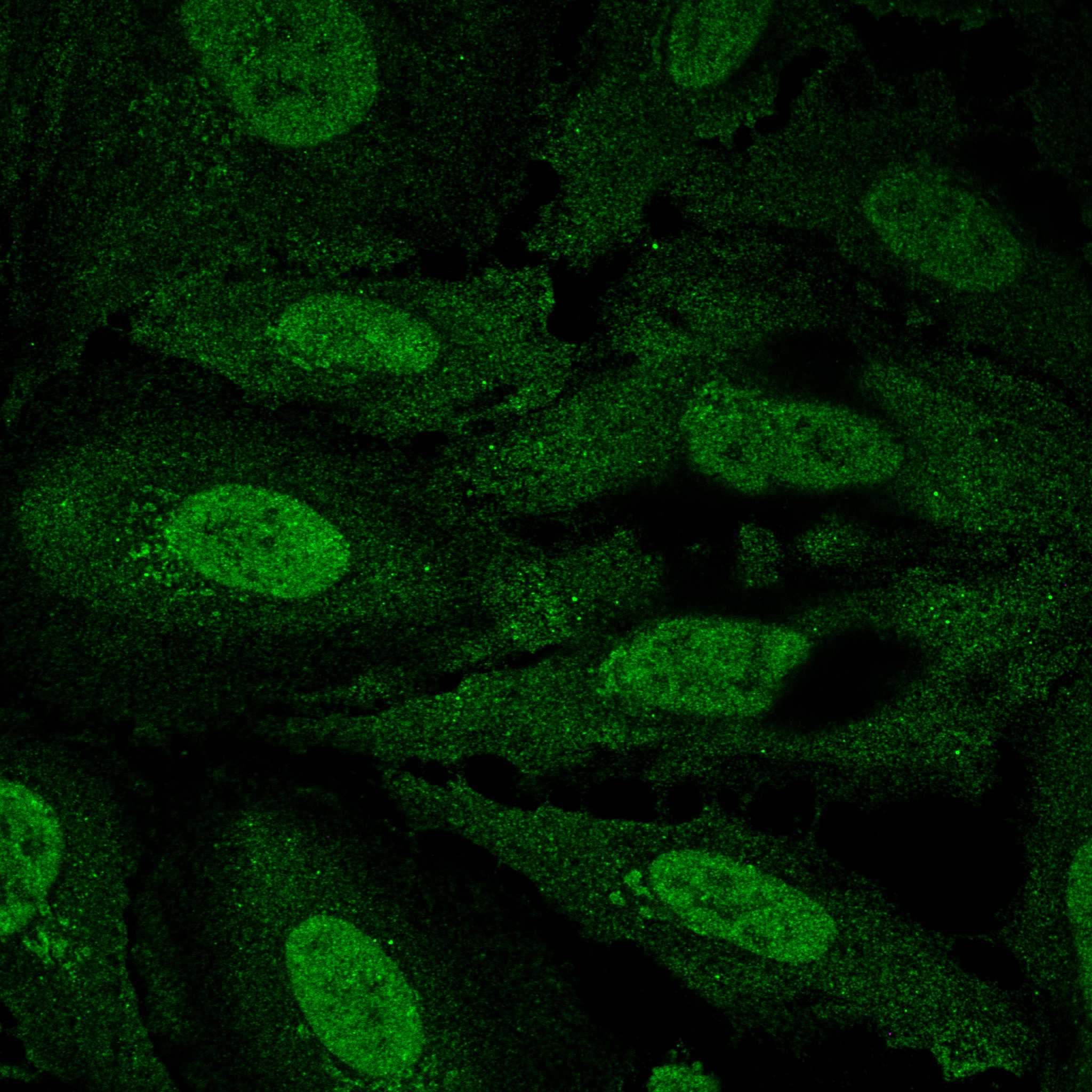
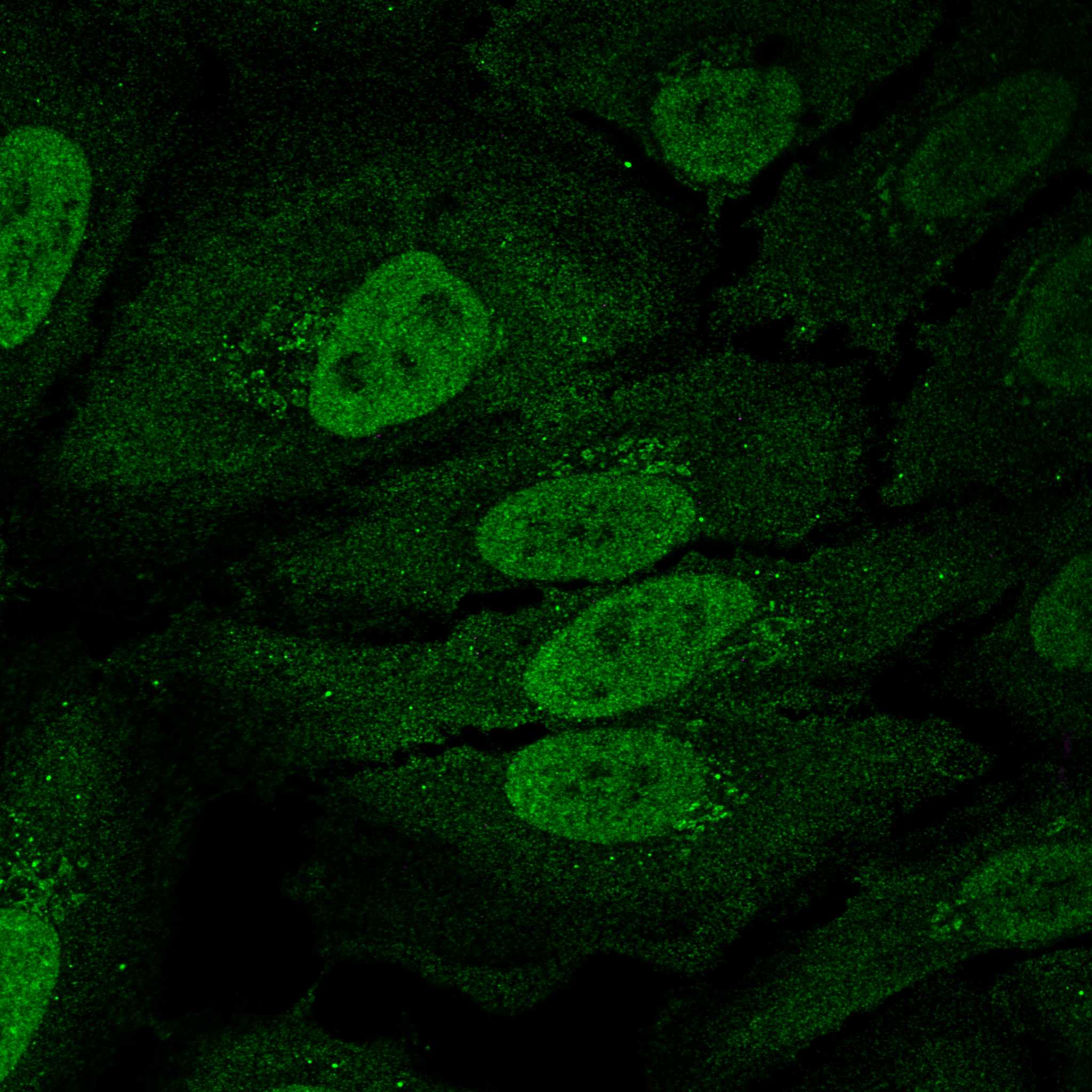
The immunofluorescent staining using the antibody in cells expressing GFP-tagged target protein is shown. The target protein can be tagged at either the N- or C-terminal and for some genes both versions are available. The antibody staining in untransfected HeLa cells is also shown. The assay is described
here.
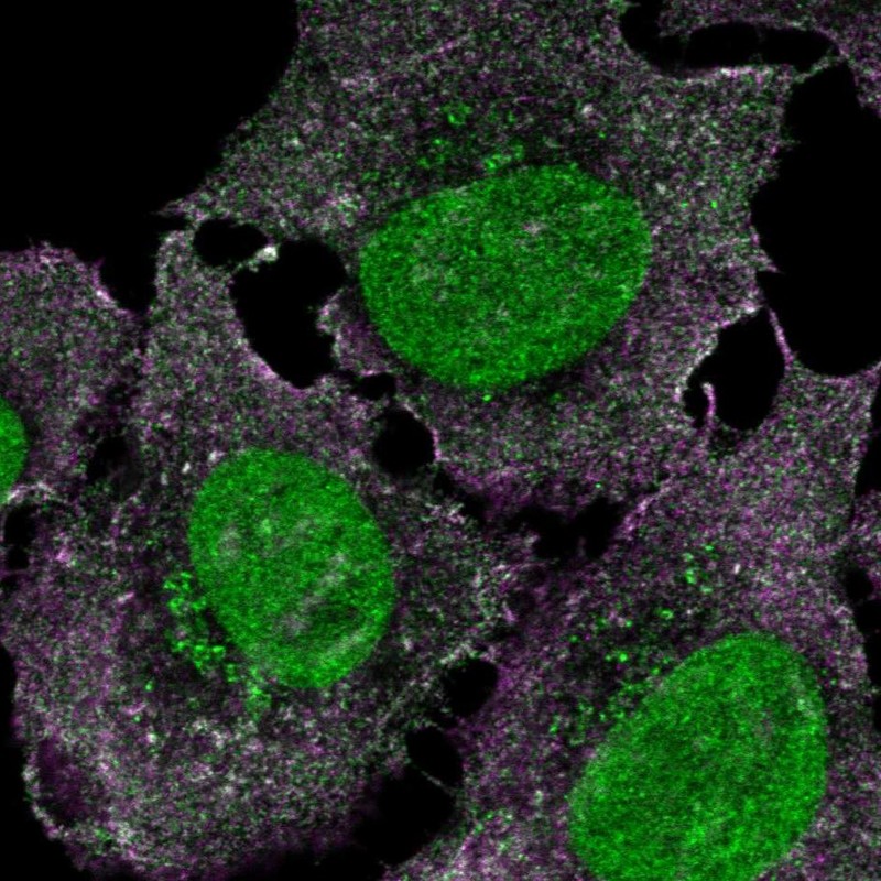
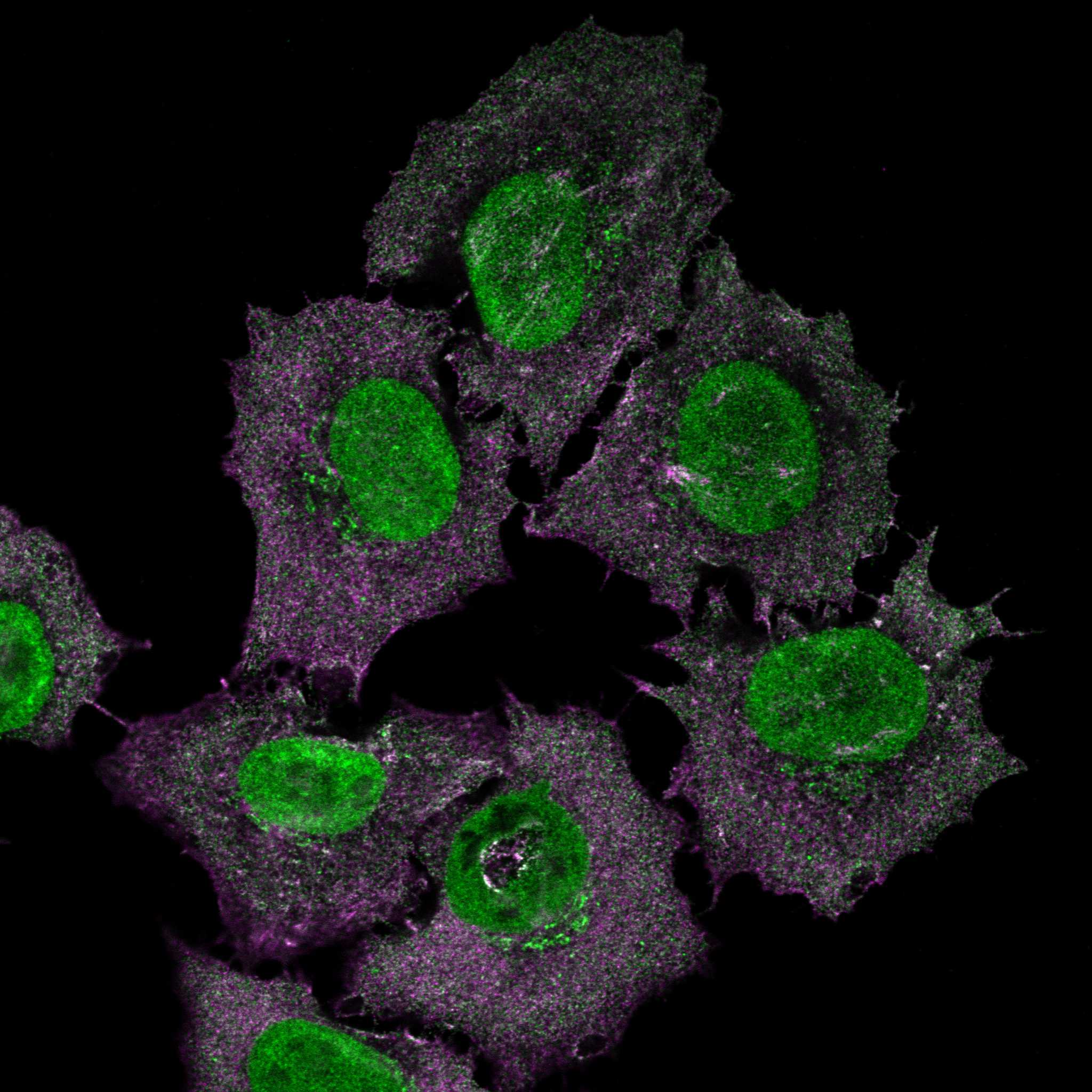
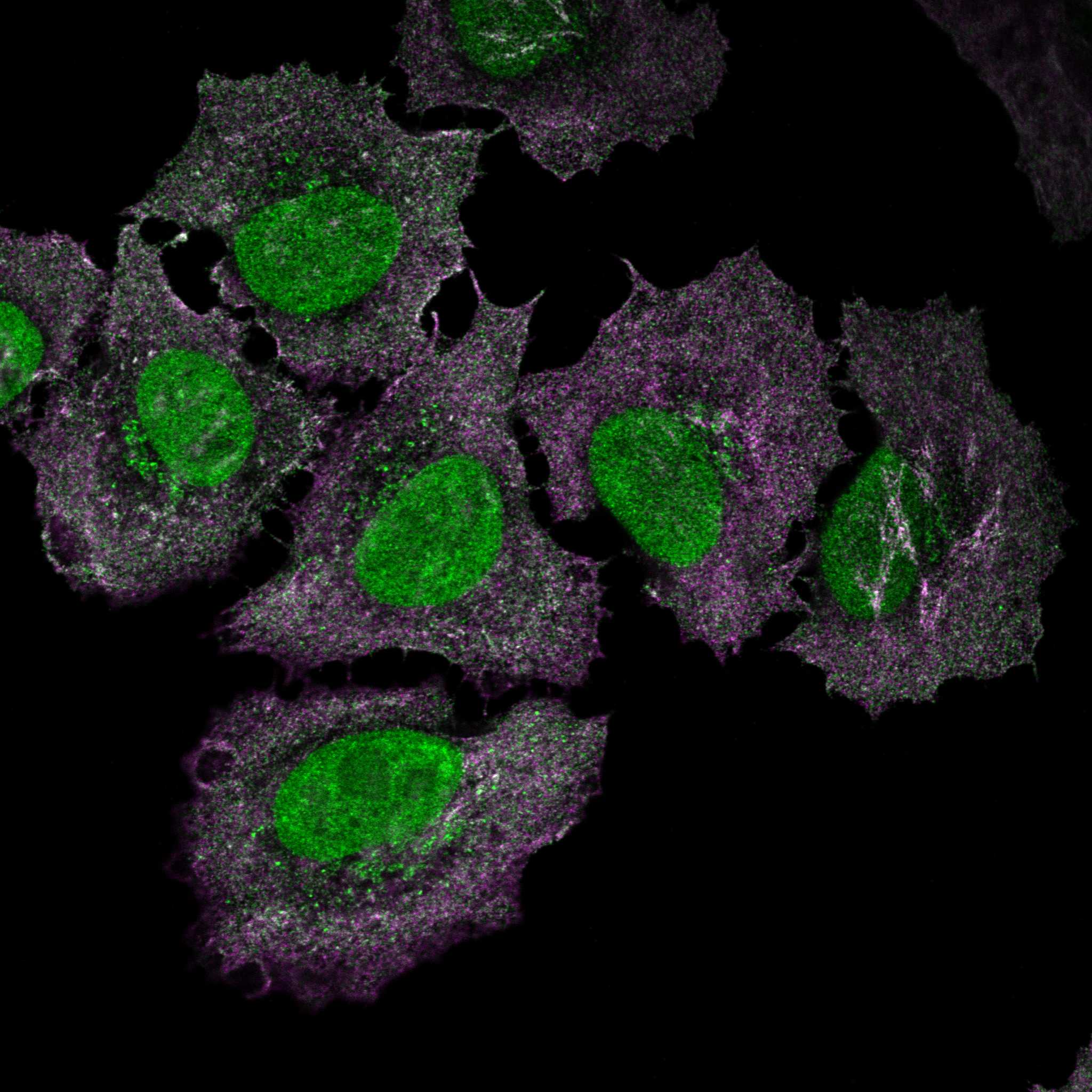
Antibody staining, GFP and two organelle probes are displayed as different channels in the multicolor images - antibody staining in green, GFP in purple, nuclear stain in blue and microtubules in red. By using the "toggle channels"-buttons, the different channels can be turned on and off. The intensity toggle shows the pixel intensity range in 16 different colors:

To change which images to compare, drag and drop a miniature image from the bottom on one of the top positions.
All images are clickable for an enlarged view. The selected image will appear in large size and miniature images with all other staining results for this gene will be listed at the top left of the image. The selected miniature image has a green overlay.
For cell structure reference, visit the
cell dictionary.